Jenn
Dearolf
CEIRP Project
As part of my CEIRP Fellowship, I work with Nancy Ridenour's 11th and 12th grade AP Biology class, and Kass Loomis' Regents Biology class, both at Ithaca High School in Ithaca, NY. I am also planning to work with Marcia Frye's Regents Biology class at Cortland High School, in Cortland, NY and Liz McCheyne's fifth grade class at South Seneca Elementary School in Interlaken, NY.
In both the
A.P. and Regents classes, we did experiments looking at cellular respiration
in muscle tissue. Both classes were studying cellular respiration,
so I designed two experiments, using techniques that are practiced
in my laboratory, for the students to complete. A description
of the basis for the activities and a summary of the labs are included
below.
I have also developed three lectures designed to introduce students to transmission and scanning electron microscopy and cellular organelles. The lectures on the transmission electron microscope (TEM) and scanning electron microscope (SEM) are designed to enhance units on microscopy. They include information about why electron microscopes can produce greater magnifications than light microscopes, how to prepare tissue for viewing on the TEM and SEM, and pictures of various specimens viewed with the microscopes. In addition, the lectures include pictures of the TEM and SEM at Cornell University.
The organelle lecture includes information about some of the most recent findings concerning the mitochondrion and the golgi apparatus. A reading from the March 5, 1999 issue of Science was given as a handout with this lecture - "Can mitochondrial clocks keep time?" by Evelyn Strauss, pages 1435-1438.
To compliment
the organelle lecture, I designed a laboratory exercise that has students
build organelles using every day items. To keep the students on task,
the exercise also includes questions they should be able to answer.
In addition, as a wrap up, the students are asked to come together
and build a cell with their organelles, and once the cell is constructed,
they discuss how different organelles communicate with each other.
Click here to download this exercise as
a PDF file, or click here to view it as
an HTML (web-based) file.
Cellular respiration experiments
Cellular respiration is a fundamental concept in biology, and many classes do laboratory exercises where students study respiration in whole organisms. This approach, although useful, cannot be used to study the different stages of cellular respiration in isolation, and it may cause students to forget that respiration is a process that occurs in the cells of the organism.
To address these two issues, I designed two laboratories using muscle tissue. Skeletal muscle is a useful tissue to study cellular respiration because its cells (fibers) have become specialized for different stages of the process. Some fibers use only glycolysis to produce the ATP needed for contraction, while other fibers utilize all of cellular respiration to fuel their activities. Thus, skeletal muscle provides a system where students can investigate the different stages of cellular respiration at the cellular level.
The first laboratory allows students to stain for the activity of an enzyme involved in the electron transport chain, nicatinamide adenine dinucleotide dehydrogenase (NADH-D). A digital image of one of the stained cross-sections of muscle is below. The fibers that are dark contain the enzyme and, thus, use all of cellular respiration to produce ATP.
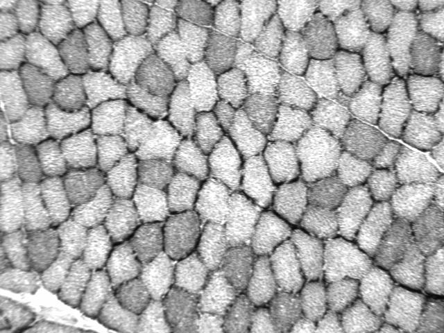
Muscle
stained for NADH-D
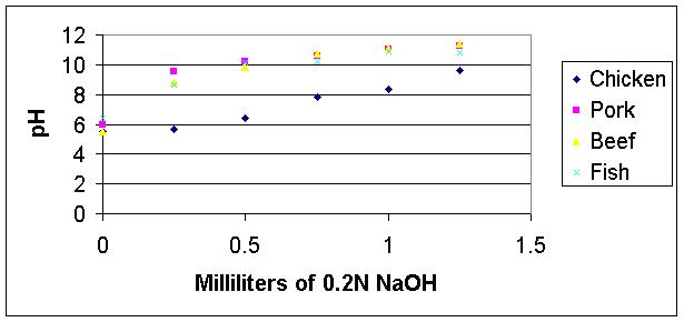
To study the fibers that utilize only glycolysis, students can investigate the acid buffering capacity of various muscle samples, like beef, pork, chicken, and fish. This investigation is based on the principle that muscle fibers that only use glycolysis to produce ATP also produce a large amount of lactic acid as a byproduct.
Thus, these cells must have some way to buffer the production of acid so that they can continue to function when lactic acid is being produced. A graph of student data is below, and it demonstrates that, out of the group of muscles investigated, the type with the greatest buffering capacity is from chicken.
Click
here for the student's manual on cellular respiration I am developing;
click here for the accompanying
teacher's manual (both in PDF format). Please note that both documents
are works in progress, and because of copyright issues, they do not
include all images and graphics.